From: Chapter 4, Atheromas Are Caseous Abscesses

NCBI Bookshelf. A service of the National Library of Medicine, National Institutes of Health.
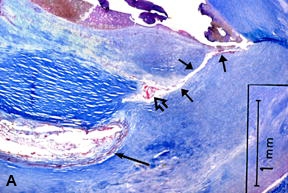
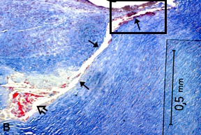
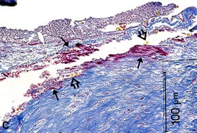
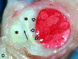

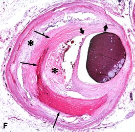
A - C, Coronary section from the proximal LAD of a 74-year-old white male. A & B, Low and intermediate power views of a plaque showing a tiny tract (arrows) traversing the fibrous cap at the shoulder, communicating with a small necrotic core, rich in fibrin (open arrow). The deeper, larger core in A (long arrow) also stains positive for fibrin, suggesting the tract also communicates with this large core. C, High-power view of rectangle shown in B at mouth of tract, showing injection mass (solid arrows) and RBCs (open arrows) within the tract. MSB stain. Fibrous tissue stains blue, fibrin orange-red, and RBCs yellow with MSB stain. D, Mid-LAD coronary artery of a 39-year-old male showing an asymmetric plaque with two lipid cores (asterisks) separated by a fibrous layer (thin arrow). Note a finger-like extension (open arrows) of one lipid core in the direction of the shoulder of the plaque, in the lower part of the photo, and thinning of the fibrous cap at this point (white arrows). The different tissue characteristics create a “layering” effect on visual examination. Magnification x12. E, Cleavage plane (arrows) in a fibrous plaque in the LAD coronary artery in a 33-year-old male. The cleavage plane contains no injection mass and is partially closed by fibrous tissue. MSB stain. F, Microscopic view of mid-LAD of a 53-year-old white female showing two atheromas (asterisks) separated by a partially calcified fibrous strand (thin arrows). Note these two atheromas are oriented toward the shoulder at the upper margin of the plaque (fat arrows). H & E stain. Magnification x19.75.
From: Chapter 4, Atheromas Are Caseous Abscesses

NCBI Bookshelf. A service of the National Library of Medicine, National Institutes of Health.