From: Chapter 1, Overview of Angiogenesis
Copyright © 2010 by Morgan & Claypool Life Sciences.
NCBI Bookshelf. A service of the National Library of Medicine, National Institutes of Health.

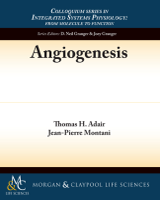
Vasculogenesis in the vertebrate embryo. (a) Angioblasts derived from lateral mesoderm are committed to become arteries (red) or veins (blue). The cardinal veins assemble from precursor cells (blue) that remain in a lateral position. (b) Artery precursor cells migrate toward a vascular endothelial growth factor type A (VEGF-A) stimulus secreted from cells in the midline. (c) The migrating arterial angioblasts align into cords forming a plexus. (d) Arterial angioblasts coalesce forming the dorsal aorta. (e) Intersomite vessels are assembled from three types of endothelial cells with different morphologies indicated as blue, purple, and green. Used with permission from Nature Publishing Group: Hogan (2002) [18].
From: Chapter 1, Overview of Angiogenesis
NCBI Bookshelf. A service of the National Library of Medicine, National Institutes of Health.