From: Chapter 1, Overview of Angiogenesis

Copyright © 2010 by Morgan & Claypool Life Sciences.
NCBI Bookshelf. A service of the National Library of Medicine, National Institutes of Health.
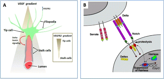
Microanatomy of a capillary sprout and tip cell selection. (A) An interstitial gradient for VEGF-A and an endothelial cell gradient for VEGFR2 are shown. Tip cell migration is thought to depend upon the VEGF-A gradient and stalk cell proliferation is thought to be regulated by the VEGF-A concentration. Redrawn after Carmeliet and Tessier-Lavigne (2005) [29]. (B) Delta-Notch signaling is critical for tip cell selection. Activation of notch receptors on stalk cells induces proteolytic cleavage and release of the intracellular domain, which enters the nucleus and decreases gene expression of VEGFR2. National Institutes of Health, public domain image.
From: Chapter 1, Overview of Angiogenesis
NCBI Bookshelf. A service of the National Library of Medicine, National Institutes of Health.