From: Chapter 1, Overview of Angiogenesis
Copyright © 2010 by Morgan & Claypool Life Sciences.
NCBI Bookshelf. A service of the National Library of Medicine, National Institutes of Health.
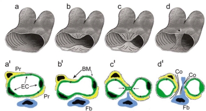
Intussusceptive angiogenesis in three dimensions (a–d) and two dimensions (a'–d'). (a,b,a',b') The process begins with protrusion of opposing endothelial cells into the capillary lumen. (c,c') An interendothelial contact is established and endothelial junctions are reorganized. (d,d') The endothelial (EC) bilayer and basement membranes (BM) are perforated centrally allowing growth factors to enter. Fibroblasts (Fb) and pericytes (Pr) migrate into the site of perforation where they produce collagen fibrils (Co) and other components of ECM forming a tissue pillar. Used with permission from Wiley-Blackwell: Djonov, Kurz, and Burri (2003) [35].
From: Chapter 1, Overview of Angiogenesis
NCBI Bookshelf. A service of the National Library of Medicine, National Institutes of Health.