From: Fusome as a Cell-Cell Communication Channel of Drosophila Ovarian Cyst

NCBI Bookshelf. A service of the National Library of Medicine, National Institutes of Health.

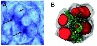
Fusome of Xenopus laevis female ovaries. A) Xenopus 8-cell cyst stained with an antibody raised against spectrin. Long arrows point to spectrin in the cytoplasm where the primary mitochondrial clouds (PMC) and the fusome localise. Short arrow indicates the nucleus. B) Three-dimensional reconstruction of an 8-cell cyst. Cytoplasm is grey, nuclei are red, mitochondria of PMC are green, centrioles blue and ring canals are yellow. PMC, ring canals and centrioles face each other and are located centripetally in the “rosette” conformation. This reconstruction was made from 38 serial ultrathin sections. (Images courtesy of Malgorzata Kloc and Laurence Etkin, Department of Molecular Genetics, University of Texas, and reprinted from: Kloc M, Bilinski S, Dougherty MT et al, Dev Biol 266:43-61, ©2004 with permission from Elsevier.8).
From: Fusome as a Cell-Cell Communication Channel of Drosophila Ovarian Cyst

NCBI Bookshelf. A service of the National Library of Medicine, National Institutes of Health.