From: Chapter 3, Inflammation. A Sign of Active Disease

NCBI Bookshelf. A service of the National Library of Medicine, National Institutes of Health.


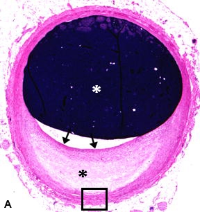
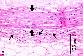
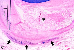
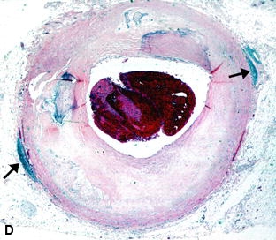
A, Proximal RCA section from a 32-year-old Asian male showing a small asymmetric plaque, with a necrotic core (black asterisk) and a fibrous cap (black arrows). White asterisk = lumen. H & E stain. Magnification x19.5. B, High-power view of rectangle in A, of the adventitia showing scattered Tcells (thin arrows). This amount of inflammatory response was classified as Grade I. Media = fat arrows. H & E stain. C, Large asymmetric atheroma (asterisk) in a 51-year-old white female with Grade II inflammation of the adventitia (arrows). Lumen at top of photo. H & E stain. D, Mid-RCA section from a 72-year-old white male. The luminal stenosis is estimated to be 80% and the lumen contains a thrombus. Two foci of T cells can be seen in the adventitia (arrows) on opposite sides of the lumen. This is classified as a Grade III inflammatory response. H & E stain. Magnification x11.2. E, Distal RCA section from a 59-year-old white male. This section was taken immediately distal to an occluding thrombus with fragments of thrombus still present in the lumen. A thick, heavy band of T cells extends virtually around the entire circumference (arrows), and this is classified as a Grade IV inflammatory response. H & E stain. Magnification x11.7.
From: Chapter 3, Inflammation. A Sign of Active Disease

NCBI Bookshelf. A service of the National Library of Medicine, National Institutes of Health.